Receiving a diagnosis of a heart blockage often leads patients and families to assume that immediate open-heart surgery or stenting is unavoidable. In contemporary clinical cardiology, however, the treatment of a heart blockage is a stepped, evidence-based process that begins long before an operating room is ever needed. Depending on whether the blockage is a mild cholesterol plaque buildup inside a coronary vessel, a severe flow-limiting arterial narrowing, a microscopic constriction of small heart vessels (Angina with Non-Obstructive Coronary Arteries – ANOCA), or an electrical signal delay (Atrioventricular Heart Block), treatment ranges from non-invasive outpatient medication and metabolic reversal to wrist-access stenting, pacemaker therapy, or coronary bypass surgery.
Across Patna and Bihar, clinicians are diagnosing coronary artery disease in a steadily younger demographic, frequently among adults in their thirties and forties. Unmanaged Type 2 diabetes, chronic hypertension, elevated low-density lipoprotein (LDL) cholesterol, tobacco consumption, and sedentary work schedules accelerate arterial plaque deposition. Catching these blockages early in an outpatient clinical setting allows cardiologists to stabilize vulnerable plaques medically—often preventing acute heart attacks and avoiding invasive procedures when narrowings do not restrict blood flow.
In strict compliance with the National Medical Commission (NMC) guidelines for factual, transparent, and non-promotional medical education, this clinical guide explains the different mechanisms of heart blockages, how non-invasive testing guides treatment decisions, and how the Ford Hospital network coordinates cardiovascular care sequentially across three dedicated facilities in Patna.
When patients use the term “heart blockage,” cardiologists evaluate three distinct physiological pathways inside the cardiovascular system. Identifying which pathway is affected determines the entire course of treatment:
Three major surface arteries—the Left Anterior Descending (LAD), the Left Circumflex (LCX), and the Right Coronary Artery (RCA), originating from the Left Main stem—supply oxygen-rich blood to the heart muscle. Over years, circulating LDL cholesterol, inflammatory cells, and calcium accumulate within the inner arterial wall to form atherosclerotic plaque. When plaque narrows the interior channel by 70% or more (or 50% or more in the Left Main trunk), blood supply falls short during physical effort, triggering myocardial ischemia. If a thin-capped lipid plaque ruptures suddenly, a blood clot can seal the artery 100% within minutes, causing an acute myocardial infarction (heart attack).
A significant subset of patients—particularly women and individuals with long-standing diabetes or hypertension—experience genuine exertional chest pain and abnormal stress tests, yet have no 70% blockages in their three main arteries. In these patients, the bottleneck occurs in the microscopic capillary branches (coronary microcirculation) that fail to dilate during exercise or undergo temporary muscular spasm. These blockages cannot be stented; they respond specifically to targeted outpatient anti-anginal pharmacology and metabolic control.
Unlike arterial blockages that restrict blood flow, an Electrical Heart Block interrupts the electrical impulses travelling from the heart’s natural pacemaker (the sinoatrial node) through the Atrioventricular (AV) node to the lower pumping chambers. While a mild First-Degree AV Block requires only outpatient medication review, a Second-Degree (Mobitz Type II) or Third-Degree (Complete Heart Block) causes the heart rate to drop below 40 beats per minute, starving the brain of oxygen and requiring a Permanent Pacemaker.
A common fear among patients is that visiting a heart specialist automatically leads to an invasive catheter test. In evidence-based cardiology, stable patients first undergo a structured 3-Step Non-Invasive Diagnostic Workup:
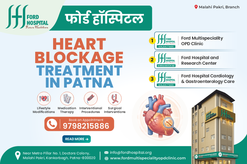
Treating heart blockages effectively requires matching the clinical setting to the exact stage and severity of the patient’s disease. Within the Ford Hospital network, care is organized sequentially across three specialized facilities in Patna: starting with our dedicated daytime outpatient clinic for early diagnosis and medical plaque stabilization, backed by our flagship 24×7 emergency and open-heart surgical campus, and supported by our dedicated interventional cardiology and gastroenterology institute.
For patients noticing early warning signs—such as mild chest tightness on walking upstairs, newly detected high blood pressure, high cholesterol, diabetic breathlessness, or a family history of premature heart disease—the Ford Multispeciality OPD Clinic serves as the primary frontline facility. Operating as a calm, crowd-free daytime outpatient center detached from emergency-room stress, this clinic currently delivers specialized OPD consultations across Cardiology, Gastroenterology, Gynecology, Psychiatry, General & Laparoscopic Surgery, Internal Medicine, and Diabetes/Endocrinology.
Clinical studies show that when coronary plaque is detected before it exceeds 50% to 60% narrowing—or when stable angina is caught early—non-invasive Guideline-Directed Medical Therapy (GDMT) can stabilize the fibrous cap of the plaque, prevent clot formation, and relieve chest pain without immediate stenting:
Outpatient Specialty Directory at Ford Multispeciality OPD Clinic:
Located near New Bypass (NH-30), Khemnichak, Patna, Ford Hospital & Research Center functions as our flagship 105-bedded multispeciality inpatient, emergency, and cardiothoracic surgical campus. When an outpatient evaluation uncovers severe multi-vessel blockages, or when a patient arrives in the middle of an acute life-threatening heart attack, this tertiary hospital provides immediate catheter, surgical, and multi-organ intensive care under one roof.
Complete 20-Department Multispeciality Directory at Ford Hospital & Research Center:
Located centrally at Malahi Pakri, Kankarbagh, Patna, Ford Hospital Cardiology & Gastroenterology Care functions as our specialized dual-specialty institute built for high-precision catheter interventions, electrical heart block devices, and the clinical co-management of overlapping heart and digestive conditions.
When a patient’s OPD stress test or symptoms indicate the need for coronary angiography or single/double-vessel stenting, this specialized center provides three distinct clinical advantages:
Explore Our Dedicated Core Services at Kankarbagh:
To help patients and family members select the appropriate Ford Hospital branch based on symptom urgency and treatment stage, review the comparison table below:
| Clinical Parameter | #1 Ford Multispeciality OPD Clinic | #2 Ford Hospital & Research Center | #3 Ford Cardiology & Gastro Care |
| Primary Role | Daytime Outpatient Screening & GDMT Hub | 24×7 Emergency, ICCU & CTVS Bypass Campus | Radial Stenting, Pacemakers & Gastro Hub |
| Location | Patna Outpatient Center | New Bypass, Khemnichak, Patna | Malahi Pakri, Kankarbagh, Patna |
| Target Blockage | Mild Plaque (under 50%) & Stable Angina | Acute Heart Attack, Triple-Vessel & Left Main | Single/Double Vessel & Electrical Block |
| Key Diagnostics | 12-Lead ECG, 2D Echo, TMT & Lipid Panels | 24×7 ECG/Troponin, Digital Cath Lab & CT | Radial Cath Lab, FFR, IVUS, OCT & Endoscopy |
| Core Treatments | Plaque Stabilization Medicines & Rehab | Primary PCI, Beating-Heart CABG & Dialysis | Complex PCI Stenting, Pacemaker & GI Care |
How to Use This Table:
Treating heart blockages safely relies on a collaborative Heart Team where clinical cardiologists, interventional specialists, and cardiothoracic surgeons evaluate cases together. In strict compliance with NMC ethical guidelines regarding transparent presentation of medical qualifications, below is our cardiac specialist directory with direct consultation links:
1. Can a heart blockage be treated with medicines alone at the OPD clinic without needing a stent or surgery?
Yes, in many cases. If diagnostic evaluation shows non-obstructive coronary plaque (less than 50% narrowing), microvascular angina (ANOCA/INOCA), or an intermediate 50% to 69% narrowing where Fractional Flow Reserve (FFR) proves blood flow is not restricted (FFR greater than 0.80), international guidelines recommend Guideline-Directed Medical Therapy (GDMT) over stenting. Using high-intensity statins, antiplatelets, anti-anginal tablets, and strict diabetes/blood pressure control at the Ford Multispeciality OPD Clinic, many patients stabilize their plaques and remain symptom-free without invasive procedures.
2. Why does a normal resting ECG not rule out a 70% to 80% coronary artery blockage?
A resting 12-lead ECG only measures electrical signals while your body is lying still and your heart muscle requires minimal oxygen. Unless you are actively having a heart attack or had a previous myocardial infarction that left electrical scar tissue, a resting ECG is often completely normal even with a 70% to 80% arterial blockage. That is why cardiologists at our OPD clinic combine an ECG with a 2D Echocardiogram and a Treadmill Stress Test (TMT)—which tests how the heart performs under physical exercise.
3. When is Angioplasty (Stenting) chosen versus Beating-Heart CABG (Bypass Surgery)?
When severe flow-limiting blockages (70% or greater) are confined to one or two coronary arteries in accessible locations, minimally invasive Radial-First Angioplasty with a Drug-Eluting Stent (PCI) is generally preferred. However, when diffuse blockages involve all three major coronary arteries (Triple-Vessel Disease) or severely narrow the Left Main stem—especially in patients with Type 2 diabetes or reduced heart pumping function (low LVEF)—Beating-Heart CABG Bypass Surgery at Ford Hospital & Research Center provides superior long-term vessel patency and survival.
4. How do FFR, IVUS, and OCT make heart blockage stenting safer?
A standard X-ray angiogram shows only a flat two-dimensional shadow of contrast dye inside the artery. FFR (Fractional Flow Reserve) measures real-time blood pressure across a borderline narrowing to confirm whether a stent is truly needed. IVUS (Intravascular Ultrasound) and OCT (Optical Coherence Tomography) pass a microscopic imaging camera inside the coronary artery to measure true vessel diameter and calcium depth, ensuring the Drug-Eluting Stent is sized accurately and expanded flush against the arterial wall—dramatically reducing the risk of future re-narrowing (restenosis).
5. Is cashless insurance available if my OPD evaluation indicates the need for angiography, stenting, or bypass surgery?
Yes. While outpatient consultations and non-invasive screening packages (ECG, 2D Echo, TMT) at the Ford Multispeciality OPD Clinic are billed transparently at standard OPD rates, if your workup indicates the need for inpatient coronary angiography, IVUS/OCT-guided angioplasty, pacemaker implantation, or CABG bypass surgery at Ford Hospital & Research Center or Ford Hospital Cardiology & Gastroenterology Care, our dedicated 24×7 TPA and insurance desks facilitate cashless approvals under major private health insurers and eligible government healthcare schemes, subject to policy terms and verification.